Bone Mineral Density (BDM)
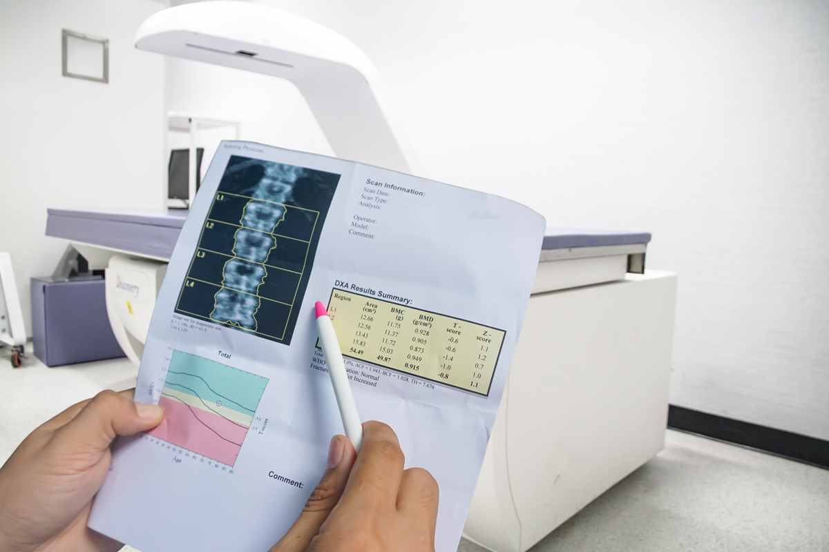
BDM scan, is a special type of X-ray that measures bone strength or fragility.
What is a bone mineral density scan?
A dual-energy X-ray absorptiometry scan (DXA), or bone density scan, is a special type of X-ray that measures bone mineral density (BMD). It provides information about bone strength or fragility and the risk of fractures or broken bones. The higher the density, generally, the lower the risk of fracture.
The spine and one or both hips are routinely scanned. The forearm might also be scanned if either the hip or spine is unavailable (usually due to surgery). As any condition affecting bone density tends to affect the whole skeleton, a snapshot of a few sites is sufficient to establish the overall bone density. The BMD at the hip and spine has been shown to be the best way of predicting the risk of fracture.
Why would my doctor refer me to have this procedure?
You might be referred for this test if you have:
-
- a medical condition that could weaken your bones;
- you might have had recent fracture after a minor injury or fall that in other persons would not have broken this bone;
- an X-ray image or picture taken for another reason has shown that the vertebrae in your spine are weakened and losing height.
The most common cause for this is osteoporosis. This is a common condition and increases with age. It is a major cause of weak bones, causing fractures resulting from minor injury, which is preventable with treatment. Osteoporosis, in the absence of fracture, has no symptoms. A number of medical conditions and medications can increase bone loss, making it important to diagnose osteoporosis early, to prevent fractures from occurring. The DXA scan measures the bone mineral content and provides information to your doctor as to whether you have lost a small amount of bone (osteopenia) or a more significant amount (osteoporosis), compared with a young normal population, and people of the same age and sex as you. This informs your doctor about your risk of having a fracture, and assists in monitoring bone loss and in planning any preventative therapy or medical treatment.
How do I prepare for a BMD scan?
No preparation is required for this procedure. You do not need to fast, and you can take all your medications as usual. There are no tunnels or confined spaces, no injections and the procedure is not painful. It is helpful, but not essential, to wear loose fitting, comfortable clothing without metal buttons, buckles, fasteners or zippers, as metal objects interfere with the scan. A gown or sheet is usually provided if clothing needs to be removed.
A DXA scan does involve a very small dose of radiation, which makes this test unsuitable for women who are, or might be, pregnant.
If you have had spinal surgery, particularly with metallic implants, or hip surgery (hip replacements, screws or pins) you will need to inform the radiographer (medical imaging technologist) carrying out the scan who might decide to avoid that area.
Any radiological investigation using contrast media (Barium enemas, IVP’s and CT scans) or nuclear medicine test might interfere with the accuracy of the DXA scan if carried out within the last week. This can usually be discussed at the time you book your DXA scan appointment.
More Information
Below are some frequently asked questions about BMD Scans.
Click on the + to read more.
What happens during a BMD scan?
On arrival for your DXA scan, your height and weight will be measured. This allows the computer to generate information about your bone density.
The most important aspect of a DXA scan is to position the hips and spine in the same way each time you are scanned, so that results are accurate and comparable at each visit. To achieve this, when scanning the spine, a cushioned box will be placed under your knees. The cushioned box allows the small of your back or lower spine to lie flat on the table. To scan the hip, this box is removed and a frame made up of a flat sheet of Perspex with a triangle at one end will be placed between your feet. The frame allows the leg being scanned to be positioned accurately. The foot is strapped to the triangle by Velcro, and the knee can also be held in place by a Velcro strap to keep the leg still. Generally, neither of these positioning manoeuvres are uncomfortable or painful.
How long does a BMD scan take?
A BMD scan varies between individual scanning machines, and can take from 10 minutes to 30 minutes.
What are the benefits of a BMD scan?
A DXA scan is currently the best test for measuring the amount of bone (density) in the spine, hip or wrist. The result is a comparison of your bone density with both the young normal population (of the same sex), and to an age- and sex-matched population. The BMD provides information about fracture risk and bone loss. It can be used to monitor response to treatment, with the usual frequency of scanning being 2 years.
More accurate assessment of BMD is obtained when the measurements are repeated at the same location and on the same machine.
Where is a BMD scan done?
BMD scanning machines are located in the radiology or endocrinology departments, or dedicated BMD units, of public or private hospitals and in private radiology practices.
Further information about a BMD scan:
Although osteoporosis is the most common cause for weak bones, there are other less common conditions that have the same effect. If your doctor is concerned about this, you might be referred for more specialised images of the bones (e.g. CT scans or X-rays), blood tests or referred to a BMD medical specialist for further advice.
Are there any after effects of a BMD scan?
There are no after effects of a BMD Scan.
What are the risks of a BMD scan?
Generally, the risks of a BMD scan by DXA are very small. The radiation doses used are extremely small and significantly less than those used in normal X-ray images, allowing the medical imaging technologist who carries out the scan to remain in the room, without the need for lead protection.
Who does the BMD scan?
A radiographer (medical imaging technologist) skilled in DXA scans will carry out the scan, and ensure you are positioned correctly and you are comfortable. In most instances the technologist will remain in the room with you for the duration of the scan.
The images taken by the technologist will be reviewed by a radiologist (specialist doctor), or another medical specialist trained in assessing the results of BMD, and a written report provided to your referring doctor.
When can I expect the results of my BMD scan?
The time that it takes your doctor to receive a written report on the test or procedure you have had will vary, depending on:
- the urgency with which the result is needed;
- the complexity of the examination;
- whether more information is needed from your doctor before the examination can be interpreted by the radiologist;
- whether you have had previous X-rays or other medical imaging that needs to be compared with this new test or procedure (this is commonly the case if you have a disease or condition that is being followed to assess your progress);
- whether or not you have had a previous BMD scan, so that the results can be compared;
- how the report is conveyed from the practice or hospital to your doctor (i.e. phone, email, fax or mail).
Please feel free to ask the private practice, clinic, or hospital where you are having your test or procedure when your doctor is likely to have the written report.
It is important that you discuss the results with the doctor who referred you, either in person or on the telephone, so that they can explain what the results mean for you.


